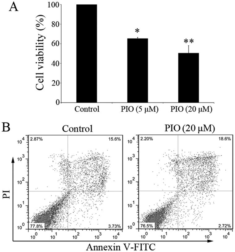
Figure 3.
PIO inhibits mast cells proliferation and induces apoptosis. (A) After 4 weeks' culture, BMMCs were starved for 6 h. Cells were cultured in complete medium with IL-3 and SCF for 7 days in the presence of PIO. Cell viability was determined by Alamar-Blue assay. The viability of control cells was set at 100% and the viability relative to control is presented. *P<0.05 vs. control, **P<0.01 vs. control. (B) BMMCs were cultured with or without PIO (20 µM) for 48 h. Cells were stained with Annexin V and PI and analyzed by flow cytometry. Data shown are representative of three independent experiments. PIO, pioglitazone; BMMCs, bone marrow-derived mast cells; SCF, stem cell factor.

