Figure 1.
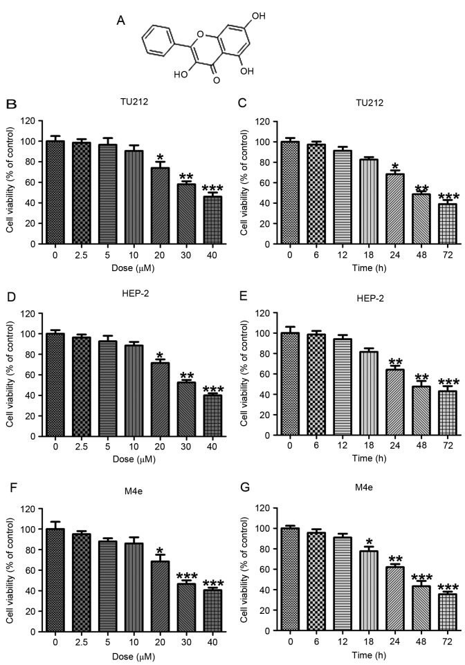
The human laryngeal carcinoma cell viability was calculated by MTT analysis. (A) The chemical structure of galangin. (B) The human laryngeal carcinoma TU212 cells were treated with different concentrations of galangin (0–40 µM) for 24 h, and the cell viability was calculated by MTT. (C) TU2I2 cells were treated with 10 µM galangin for the indicated time. The cell viability was evaluated by MTT assays. (D) The human laryngeal carcinoma HEP-2 cells were treated with different concentrations of galangin (0–40 µM) for 24 h, and then the cell viability was calculated by MTT. (E) HEP-2 cells were treated with 10 µM galangin for the indicated time. The cell viability was evaluated by MTT assays. (F) The human laryngeal carcinoma M4e cells were treated with different concentrations of galangin (0–40 µM) for 24 h, and the cell viability was calculated by MTT. (G) M4e cells were treated with 10 µM galangin for the indicated time. The cell viability was evaluated by MTT assays. All analysis were conducted in triplicate, and the results are the mean ± SEM of three independent experiments. *P<0.05, **P<0.01 and ***P<0.001 (compared to the control/Con).

