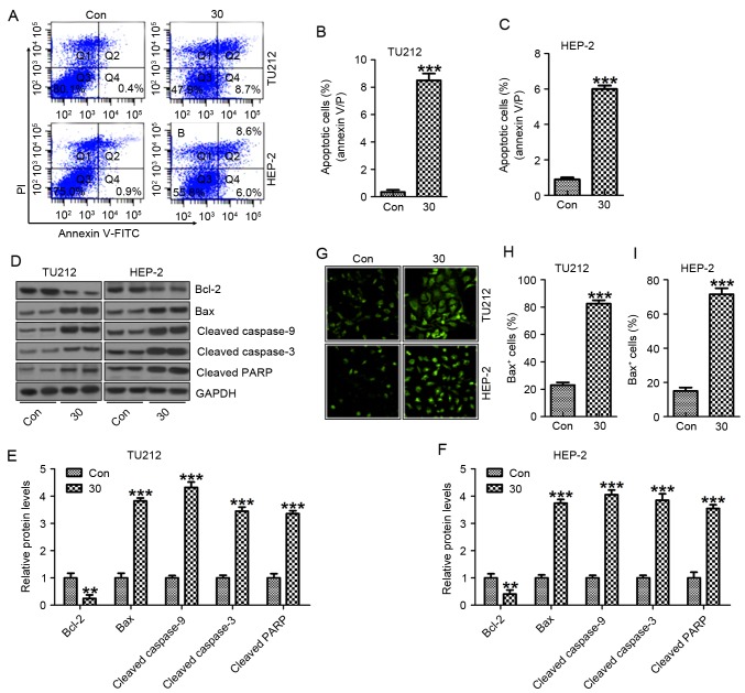
Figure 5.
Galangin induces apoptosis in human laryngeal carcinoma TU212 and HEP-2 cells. (A) Flow cytometric assays were applied to determine the number of apoptotic TU212 and HEP-2 cells. (B) The quantification of TU212 apoptosis levels is shown. (C) The quantification of HEP-2 apoptosis levels was shown. (D) The signaling pathway, leading to apoptosis, was analyzed by western blot analysis in TU212 and HEP-2 cells. Protein levels of Bcl-2, Bax, cleaved caspase-9, cleaved caspase-3 and cleaved PARP were evaluated. (E) Preotein levels of Bcl-2, Bax, cleaved caspase-9, cleaved caspase-3 and cleaved PARP in TU212 cells were quantified. (F) Protein levels of Bcl-2, Bax, cleaved caspase-9, cleaved caspase-3 and cleaved PARP in HEP-2 cells were quantified. (G) Immunofluorescence assays were carried out to determine Bax positive TU212 and HEP-2 cells. The quantification of Bax positive (H) TU212 and (I) HEP-2 cells are displayed. The analysis was conducted in triplicate, and the results exhibit the mean ± SEM of three independent experiments. **P<0.01 and ***P<0.001 (compared to the control/Con).

