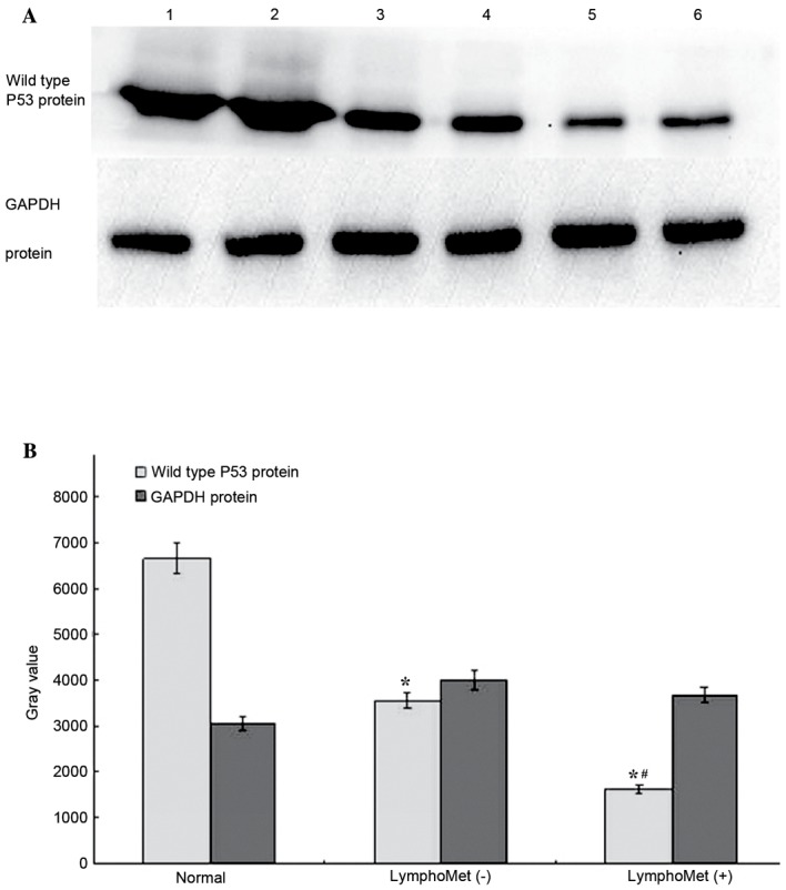
Figure 7.

(A) Western blot analysis to measure the TP53 protein expression in 30 cases of ESCC with lymph node metastasis, 30 cases without and 10 cases of normal esophageal epithelial tissues. (B) Both the tumors with metastasis and the tumors without metastasis had significantly reduced TP53 protein expression compared to the normal tissues (~75.5%, 46.7% reduction, respectively). The tumors with metastasis had further reduced TP53 expression compared to the tumors without metastasis (~54.1% reduction). Data are presented as means ± standard deviation. *P<0.05 vs. normal group; #P<0.05 vs. LymphoMet (−) group.
