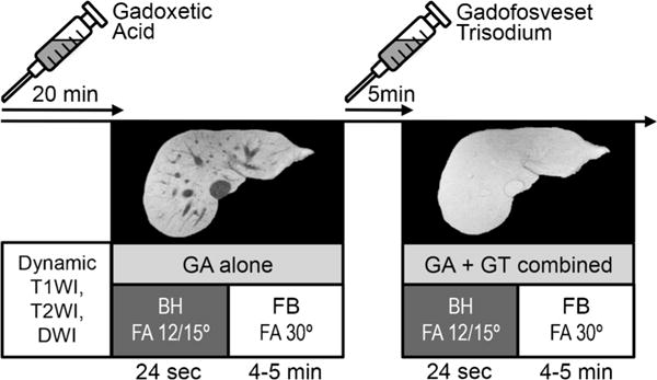
Fig. 2.

Overview of the clinical protocol. Twenty minutes after injection of gadoxetic acid, breathheld (BH) and respiratory-gated free-breathing (FB) T1-weighted imaging was performed at 12° (1.5 T) or 15° (3 T) and 30° (both 1.5 and 3 T) flip angle, respectively. Five minutes after injection of gadofosveset trisodium, imaging was repeated with identical imaging parameters during the steady-state blood pool phase of gadofosveset while still in the hepatobiliary phase of gadoxetic acid. T1WI T1-weighted imaging, T2WI T2-weighted imaging, DWI diffusion-weighted imaging, GA gadoxetic acid, GT gadofosveset trisodium, FA flip angle. Note the isointensity of the vessels relative to the liver tissue after injection of gadofosveset
