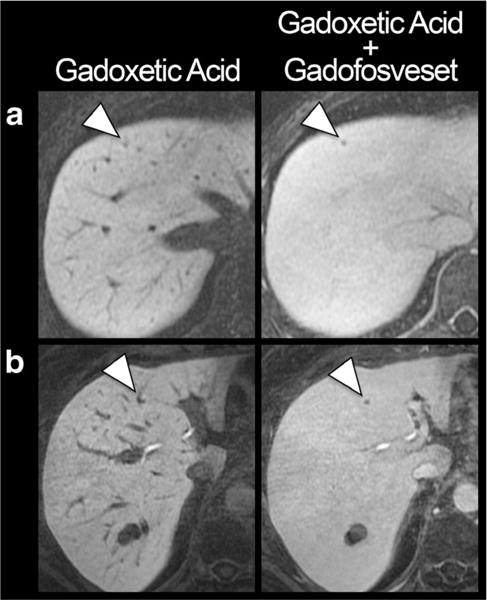
Fig. 3.

Improved conspicuity of small liver metastases. T1-weighted images with gadoxetic acid alone and in combination with gadofosveset in a 41-year-old woman with melanoma (a) and a 53-year-old woman with oesophageal cancer (b). Note the improved conspicuity of the metastases (arrowheads), particularly of the perivascular lesion in b. Both histologically confirmed metastases were missed by both readers on gadoxetic alone enhanced images but detected on combined gadoxetic acid/gadofosveset-enhanced images. The two patients had only one and two other metastases, respectively
