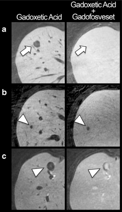
Fig. 5.

Different enhancement patterns of haemangiomas and metastases. All lesions appear hypointense compared the liver tissue on gadoxetic acid alone enhanced liver MRI. a Example of a 50-year-old woman with haemangioma (arrow). The additional injection of gadofosveset in the hepatobiliary phase of gadoxetic acid renders the haemangioma isointense to liver tissue. b Example of a 58-year-old woman with a metastases of colorectal cancer, showing no or negligible enhancement after injection of gadofosveset (arrowhead). c Example of a 51-year-old man with metastases of a neuroendocrine carcinoid tumour, showing central enhancement after injection of gadofosveset (arrowhead)
