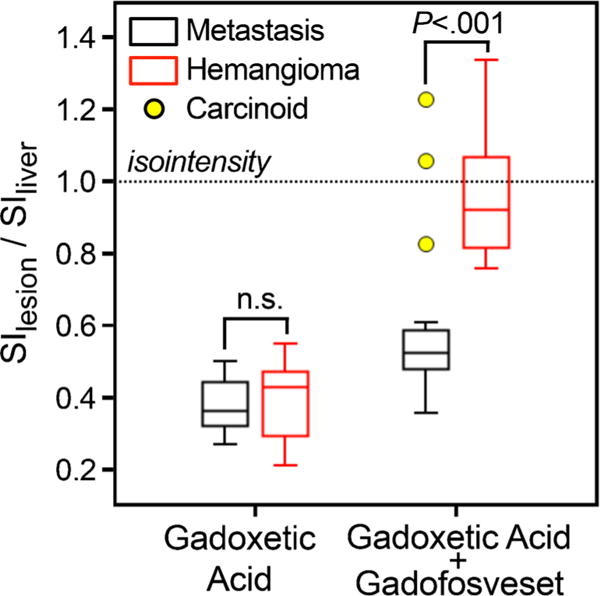
Fig. 6.

Improved differentiation of metastases vs. haemangiomas with combined gadoxetic acid and gadofosveset-enhanced liver MRI. Using gadoxetic acid alone does not allow differentiation of metastases from haemangiomas by signal intensity (P = 0.51); both appear hypointense and have low SI ratios relative to the liver. Injection of gadofosveset leads only to a slight increase of the liver/metastasis SI ratio, whereas the haemangioma/liver SI ratio is dramatically increased, thereby rendering the SI ratios of haemangiomas and metastases significantly different (P<0.001). Dotted horizontal line indicates isointensity of lesions with liver tissue. Outliers (yellow circles) represent contrast-enhancing metastases in a patient with carcinoid tumour. Note, however, that the enhancement pattern (central) of these carcinoid metastases made it easy to distinguish all of these lesions from haemangiomas (see Fig. 5)
