Fig. 3.
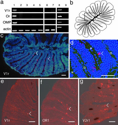
Expression of V1r-like transcripts in the zebrafish olfactory system. (a) Expression of Dr V1r mRNA. cDNA from different adult tissues was amplified by PCR with specific primers for DrV1r, odorant receptor 1 (OR), olfactory marker protein (OMP), and actin. Lane 1, olfactory rosette; lane 2, gills; lane 3, olfactory bulb; lane 4, brain; lane 5, heart; lane 6, barbels; lane 7, lips; lane 8, genomic DNA; lane 9, olfactory rosette minus reverse transcriptase. Because no introns are found in the coding sequence of V1r and OMP, the sizes of the cDNA amplicons correspond to the size of the genomic amplicons. The amplicons corresponding to actin amplified from genomic material contain an intron and are therefore larger than cDNA amplicons. Exclusive V1r expression is observed in the olfactory rosette. (b) Drawing of a horizontal section of an olfactory rosette (lamellae are cut perpendicular to their flat face). The gray zone in the lamellae, close to the center, indicates the location of the sensory neuroepithelium. The black lines correspond to the cartilage. The rectangle indicates the area shown in d. (c and d) In situ hybridizations of horizontal sections through the olfactory rosette, with an antisense DrV1r-like RNA probe. Neurons expressing V1r-like mRNA appear as fluorescent red. The blue color corresponds to DAPI staining of the nuclei. The asterisks and squares indicate the lumen between the lamellae and the extracellular matrix, respectively. (e–g) In situ hybridizations with V1r, OR1, and V2r1 probes, respectively. The numerous neurons positive for V2r1 likely reflect crosshybridization with multiple V2r sequences. (Scale bar, 30 μm.) Arrowheads indicate some of the labeled neurons.

