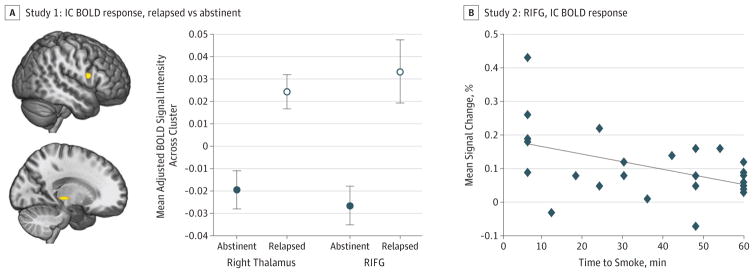
Figure 1. Associations Between Inhibitory Control (IC) Blood Oxygenation Level–Dependent (BOLD) Response and Smoking Outcomes.
A, In study 1 (cessation study), abstinent smokers exhibited less baseline (ie, prequit attempt) IC task-related BOLD response in the right inferior frontal gyrus (IFG) (yellow cluster in top figure) and right thalamus (yellow cluster in bottom figure) than did smokers who relapsed (P<.001 cluster-determining threshold, cluster extent >186 mm3; 3dClustSim autocorrelation function) (eTable 3 in the Supplement). Error bars represent 1 SE. B, In study 2 (laboratory study), greater BOLD response in the right IFG during IC predicted a shorter time to smoke during the smoking relapse analog task (SRT) (β = −0.468; P = .02; adjusted R2 = 0.151) (eTable 3 in the Supplement). All imaging data were analyzed within an IC network mask (eFigure 2 in the Supplement).

