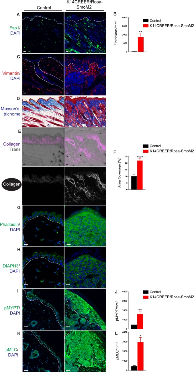
Fig 2. Activated ROCK-signalling, increased dermal fibroblast numbers, and dermal fibrosis in the skin of K14-CreER/Rosa-SmoM2 transgenic mice.
(A-G) Immunofluorescence staining and area coverage analysis of dorsal skin tamoxifen- and vehicle-treated K14-CreER/Rosa-SmoM2 transgenic littermate mice detecting Fsp1 (A & B) and Vimentin (C), Phalloidin (G), DIAPH3 (H), Thr696-phosphorylated MYPT1 (I & J), Thr18/Ser19-phosphorylated MLC2 (K & L). (D) Masson’s trichrome histological staining of sections through the dorsal neck skin of tamoxifen- and vehicle-treated K14-CreER/Rosa-SmoM2 mice. (E & J) Dual two-photon SHG and monochromatic transmission (Trans; grayscale in merge) images showing collagen (white in single channel, magenta in merged) in tamoxifen- and vehicle-treated K14-CreER/Rosa-SmoM2 skin sections. Area coverage analysis (5 fields/sample from three mice per genotype) of SHG is quantified. Basement membranes and hair follicles are demarcated with dashed lines. DAPI, 4, 6-diamidino-2-phenylindole. Scale bars = 20 μm.

