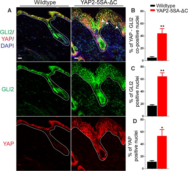
Fig 3. GLI2 activation in the skin of YAP2-5SA-ΔC transgenic mice.
(A) Immunofluorescence staining of dorsal skin sections of YAP2-5SA-ΔC transgenic and wildtype mice detecting GLI2 (green) and YAP (red). Quantification of % YAP-GLI2 co-positive (arrowheads—B), % GLI2 positive (C) and % YAP (D) positive nuclei in the skin sections of YAP2-5SA-ΔC transgenic and wildtype mice. Basement membranes are demarcated with dashed lines. DAPI, 4, 6-diamidino-2-phenylindole. Scale bars = 20 μm.

