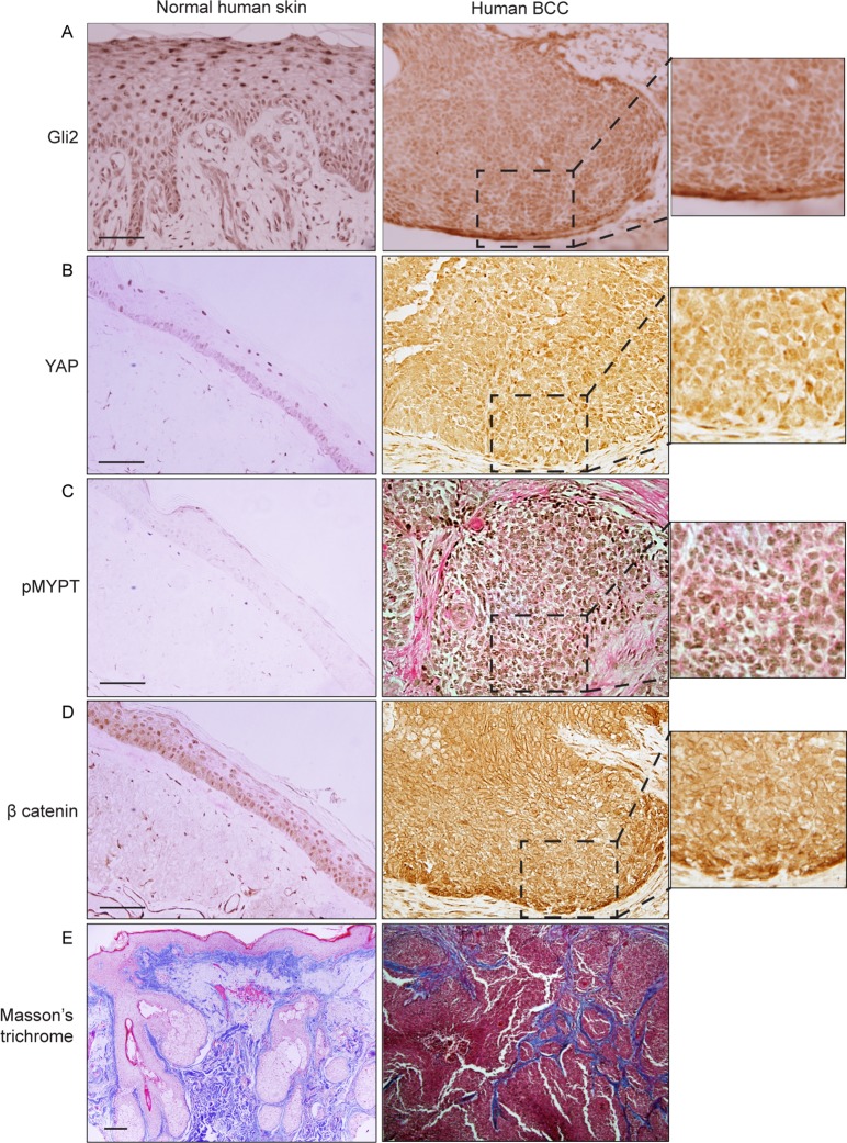
Fig 5. Human BCCs exhibit nuclear YAP and β-catenin in association with ROCK signalling activation and increased ECM collagen deposition.
Representative images of immunohistochemical staining (brown) of Gli2 (A), YAP (B), Thr696-phosphorylated MYPT (C) and β-catenin (D) in normal and human BCCs skin samples. (E) Masson’s trichrome histological staining. IHC, Immunohistochemistry. Scale bars = 20 μm.

