
Figure 4.
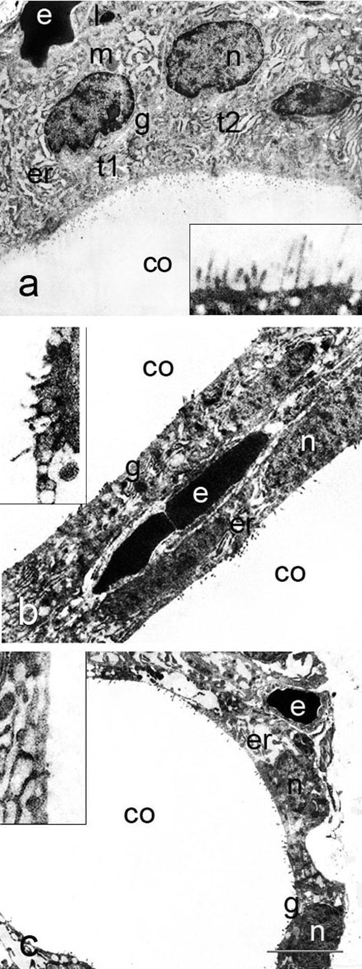
Follicular epithelium of control, T3- and T4-treated rats (a, b and c); t – thyrocyte, n – nucleus, m – mitochondrion, l – lysosome, g – Golgi complex, er – endoplasmic reticulum, co – colloid, e – erythrocyte in the capillary lumen. Insets show apical pole of thyrocytes, with reduced and disorganized microvilli in T3- and T4-treated rats. Bar 4 μm (a–c) and 1.5 μm (insets).