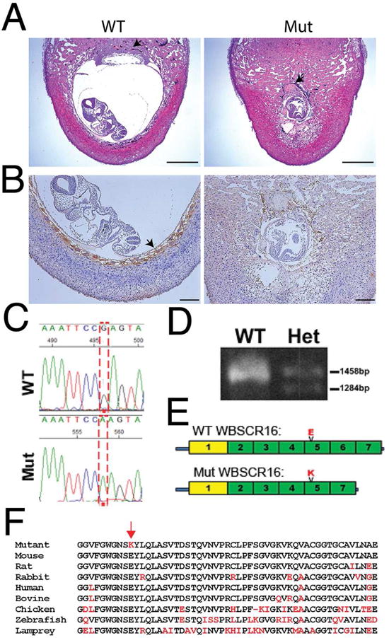
Figure 1. A Wbscr16 mutation causes early embryonic lethality.

(A) H&E stained sections of implantation sites for Pcolce−/− E8.5 embryos found to be wild type (WT) or homozygous for a mutation (Mut), at the Wbscr16 locus. Both embryos were on a C57BL/6 background. Black arrows; ectoplacental cone (WT), fibrin/platelet clot replacing the ectoplacental cone (mutant). Scale bars 600 μm. (B) WT and mutant sections were immunostained (brown) for trophoblast giant cell (TGC) marker placental lactogen 1 (PL-1). Arrow, HRP signal for anti-PL-1 (WT). Brown color in mutant is mostly due to blood cells, but also to sparse HRP-stained TGCs with abnormally small nuclei. Scale bars 50 μm. (C) G to A transition (reverse strand). (D) PCR-amplified cDNA shows normal size Wbscr16 mRNA, plus a 164 base-smaller mRNA in heterozygotes not detected in WT. (E) Mutant WBSCR16 p.E308K substitution in RLD 5 and loss of RLD 6 by alternative splicing. Green, canonical RLDs; yellow, non-cannonical RLD. (F) RLD 5 aligned across vertebrate species (black, conserved residues; red, residues that do not match WT murine sequence). Red arrow, mutant Lys that substitutes for highly conserved Glu. See also Figures S1 and S2.
