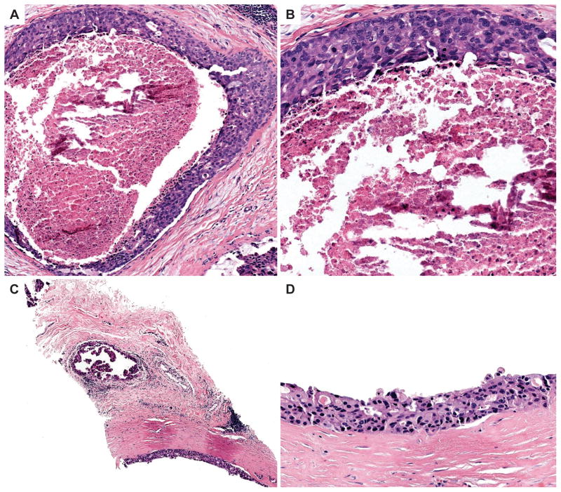
Figure 3.
A–B) The top two panels show 10x (A) and 20x (B) images from a case with 100% diagnostic agreement with the consensus diagnosis of high grade DCIS. There is obvious central comedo-necrosis present in the center of a duct lined by a proliferation with pleomorphic, hyperchromatic nuclei. C–D) The bottom two panels show 10x (A) and 20x (B) images of an unusual case that had very low diagnostic agreement (7%) with the consensus high-grade DCIS diagnosis. This case lacks necrosis and the nuclei of the proliferation are not as obviously high grade as those shown in A–B. A second duct contains micropapillary structures with a similar cytology (B). The majority of participants recorded a highest order diagnosis of ADH (88%) for this case.

