. 2017 Aug 18;6:e27301. doi: 10.7554/eLife.27301

Figure 3. LRP16 is required for genotoxicity-induced NF-κB activation.

(A) Whole cell lysates from cells transfected with an LRP16-expressing plasmid or the control vector were treated with the indicated concentrations of etoposide for 2 hr or with 50 μM etoposide for the indicated periods, and then immunoblotted with the indicated antibody. β-Actin was used as the loading control. (B) SW480 cells transfected with an LRP16-expressing plasmid or the control vector were treated with IR for the indicated dose. The cell lysates were then separated with SDS-PAGE and analyzed with Western blotting using the indicated antibody. (C) Whole-cell lysates from SW620 cells transfected with the indicated siRNAs and treated with the indicated concentrations of etoposide for 2 hr or with 50 μM etoposide for the indicated periods were immunoblotted with the indicated antibody. β-Actin was used as the loading control. (D) SW620 cells were transfected with the indicated siRNAs and exposed to IR for the indicated periods. Their lysates were separated with SDS-PAGE and analyzed with Western blotting using the indicated antibody. (E) SW620 cells were cotransfected with the indicated siRNAs and/or the LRP16-expressing vector that contained silent mutations in the sequences that targeted by the LRP16 siRNAs, and were then treated with 50 μM etoposide for the indicated periods. The cell lysates were immunoblotted using the indicated antibody. (F) Whole cell lysates of cells transfected with the control vector or vectors expressing LRP16 or the LRP16 mutants (LRP16_D160A, LRP16_I161A) and treated with the indicated concentrations of etoposide for 2 hr or with 50 μM etoposide for the indicated periods were immunoblotted with the indicated antibody. β-Actin was used as the loading control. One representative experiment of three was shown.

Figure 3.

Figure 3—figure supplement 1. LRP16 executes an essential function in the nuclear-initiated NF-κB signaling in response to DNA damage.

Figure 3—figure supplement 1.

(A) LoVo cells transfected with control vector or LRP16-expressing plasmid were treated with the indicated concentrations of etoposide or with etoposide for the indicated periods, and their lysates were separated with SDS-PAGE and analyzed with Western blotting using the indicated antibody. β-Actin was used as the loading control. (B) LoVo cells transfected with control vector or LRP16-expressing plasmid were treated with the indicated concentrations of etoposide or with the indicated doses of IR for 2 hr, and their lysates were analyzed with Western blotting using the indicated antibody. β-Actin was used as the loading control. (C) Cytosolic and nuclear fractions derived from SW480 cells transfected with an LRP16-expressing plasmid or the control vector for 48 hr and then treated with etoposide (50 μM) for the indicated periods were immunoblotted with the indicated antibody. β-Tubulin and Sp1 were used as the loading controls and the cytosolic and nuclear markers, respectively. (D) SW620 cells were transfected with the indicated siRNAs and exposed to 50 μM etoposide for the indicated periods. The cytoplasmic and nuclear fractions were then prepared and immunoblotted with the indicated antibodies. One representative experiment of three was shown.
Figure 3—figure supplement 2. LRP16 is required for NF-κB transactivation in response to genotoxic stress.

Figure 3—figure supplement 2.

(A) NF-κB transcriptional activity. SW480 cells or LoVo cells were co-transfected 3 × κB-luc luciferase reporter constructs with the indicated plasmids for 48 hr followed by the treatment with etoposide or IR. Luciferase activity was normalized by that in the vehicle group. (B) SW620 cells or HCT116 cells co-transfected 3 × κB-luc luciferase reporter constructs with the indicated siRNAs for 48 hr were treated with etoposide or IR for luciferase activity assay. Error bars in (A) and (B) represent the mean ± SD for triplicate experiments. *p<0.05, **p<0.01.
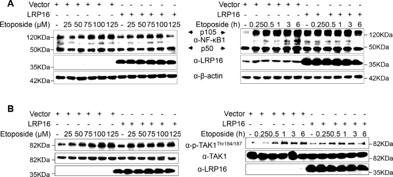
Figure 3—figure supplement 3. LRP16 is not critical for NF-κB1 and TAK1 activation during the cellular response to gentoxic stress.

Figure 3—figure supplement 3.

(A–B) SW480 cells transfected with control vector or LRP16-expressing plasmid were treated with the indicated concentrations of etoposide or etoposide for the indicated periods. Their lysates were subjected to immunoblotting using the indicated antibody. One representative experiment of three was shown.
Figure 3—figure supplement 4. Ectopic of LRP16 augments DNA-damage-triggered NF-κB-mediated expression of anti-apoptotic molecules.

Figure 3—figure supplement 4.

(A) The expression of different genes in SW480 cells that were transfected with an LRP16-expressing plasmid or the control vector followed by the treatment with etoposide were assessed with a gene expression microarray and is presented as a heat map. (B) Analyses of KEGG and GO functional pathway were performed using the gene expression microarray data. (C) RT–qPCR measurement of the expression of the indicated genes, selected from the PCR array results for human NF-κB signaling pathway, in SW480 cells during the ectopic expression of LRP16 or the control cells followed by the treatment with etoposide (50 μM) for 1 hr. One representative experiment of three was shown.