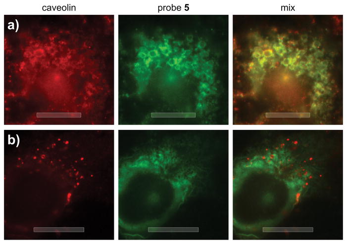
Figure 5.
Epifluorescence imaging of the uptake of probe 5. U2OS cells were incubated with 50 μM 5 in DMEM buffer for 10 min. After treatment, the cells were fixed and stained with 1:200 dilution anti-CAVEOLIN mAb (D46G3, Cell Signaling) and 80 μM Alexa647-conjugated anti-IAF mAb (XRI-TF35, Xenobe Research Institute). Localization of CAVEOLIN (red) was completed by staining with a Cy3B-labeled secondary antibody and imaging with λex 535–585 nm and λem 600–660 nm. The localization of IAF tag (green) was conducted by imaging with λex 590–650 nm and λem 663–738 nm. b) Comparable image collected after incubation with 50 μM 5 for 75 min indicating complete localization in the ER. Bars denote 10 μm.

