
Figure 5.
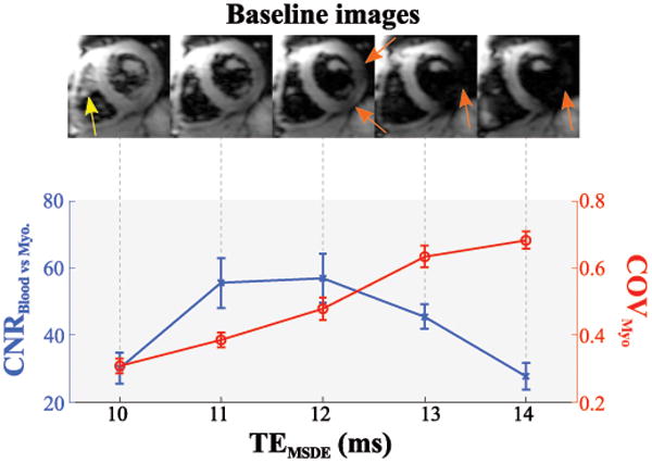
Black-blood contrast as a function of the MSDE preparation echo time. Exemplary baseline images (upper panel) show residual blood-signal for too short echo-times (yellow arrow), while long echo-times cause myocardial signal void (orange arrows). Accordingly, the contrast-to-noise ratio (CNR) between myocardium and blood-pool is compromised for long and very short echo-times. The coefficient of variation (COV) in the myocardium increases with longer echo-times, caused by progressive signal void.