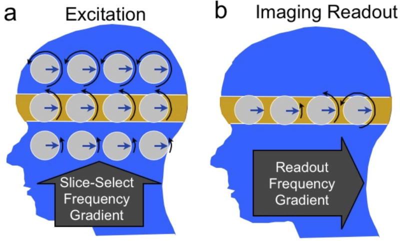
Figure 2.
Magnetization dynamics during slice selection (a) and imaging readout (b). The magnetization precession rate or frequency is indicated by black arrows showing different amounts of phase (rotation). To excite the highlighted slice (a), a gradient is turned on in the slice-select direction (superior-inferior) to impose a frequency variation. A radiofrequency (RF) pulse excites the magnetization at the frequency of the desired slice. Once the slice has been excited (b), the slice-select gradient is turned off, and a gradient is turned on in the readout or “frequency-encode” direction (anterior-posterior). This imposes a frequency variation with position. The received signal then consists of multiple frequencies, the strength of which depend on the amount of signal at the corresponding position.

