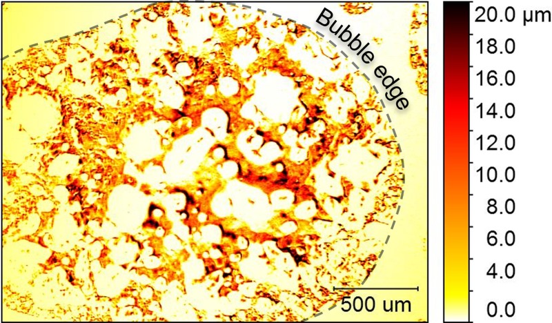
FIG. 5.
Insertion of a bubble creates an air/water interface that made it possible to use WLI to image the biofilm structure. This structure was only revealed under the air bubble and not beyond the edges of the bubble (indicated by a dashed line). False coloring in the image corresponds to biofilm thickness, with darker regions indicating a thicker growth of the biofilm.

