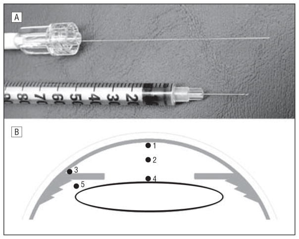
Figure 1.
Instrument and technique of PO2 measurement in the human eye in vivo. A, Optical oxygen sensor shown next to a standard 30-gauge tuberculin syringe needle. B, The locations in the eye where PO2 was measured: (1) beneath the central corneal endothelium, (2) mid–anterior chamber, (3) anterior chamber angle, (4) anterior lens surface, and (5) posterior chamber.

