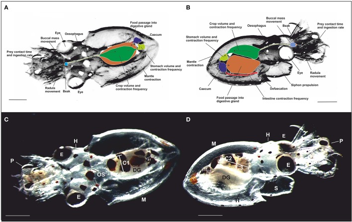
Figure 1.
Schematic representation of the dorsal (A) and the ventral (B) view of a paralarva in contact with prey (crab zoea) showing the location of the main digestive tract structures measured. Arrows indicate the direction of the food passage. (C,D) photography of the dorsal view (A) and the ventral view (B) taken from video recordings of a 3 dph free-swimming paralarvae of O. vulgaris with crab zoea prey captured. Note the transparency of the paralarva enabling direct observation of the digestive tract. The diameter of the crop (C) and the stomach (S) was measured along three axes: C1 and S1 (medio-lateral width), C2 and S2 (rostro-caudal length), and C3 and S3 (dorso-ventral thickness). OS, Oesophagus; DG, digestive gland; TI, terminal part of the intestine; E, eye; H, head; P, prey; M, mantle; and S, siphon. Scale bar = 0.5 mm.

