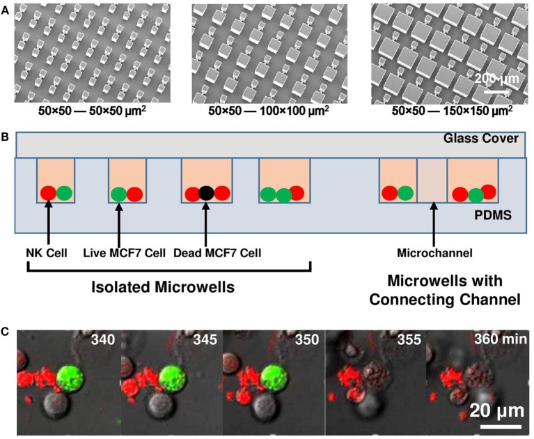
Figure 1.
Experiment setup. (A) Micrographs of SU-8 stamps to duplicate polydimethylsiloxane (PDMS) platforms. (B) Schematic of microwell and microchannel arrays with natural killer (NK) cells (red) and MCF7 cells (green: live; black: dead) loaded. (C) Sequence of MCF7 cell lysis as MCF7 cell lost its green fluorescent protein signal at 355 min.

