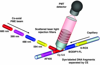
Fig. 1.
Drawing of the PME technology. Here, each laser operates in a continuous-wave mode with mechanical shutters pulsing the different excitation beams in sequential order. An arrangement of steering mirrors directs the pulsed laser beams into a dual prism assembly, where the prisms are positioned at a 45° angle relative to one another. This allows efficient convergence in overlapping the four beams into a single beam by the process of inverse dispersion. The single coaxial PME beam then interrogates the fluorescently labeled DNA fragments, which are separated by capillary gel electrophoresis. Scattered laser light is rejected via specific long-pass or wavelength notch filters, with pulsed emission signals from the dye-labeled DNA fragments being imaged onto the photomultiplier (PMT) detector without use of any dispersing elements. The fluorescence is detected directly from the cuvette in an orthogonal geometry (not shown) or from the capillary, which is positioned 45° relative to the optical axis of the detection train to further reduce scattered laser light.

