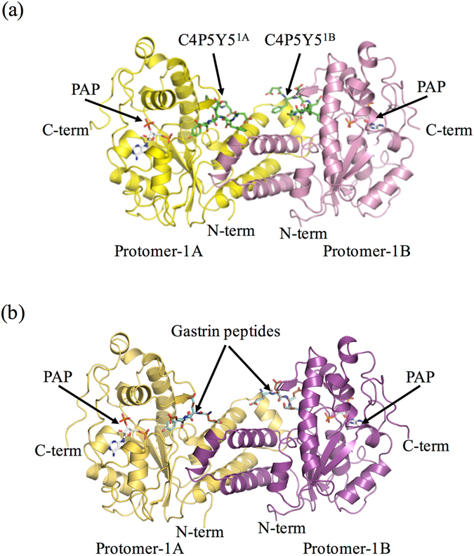
Figure 2.

Overall structures of recombinant human TPST1. (a) Ribbon diagram of the structure of human TPST1-PAP-C4P5Y5. Protomer-1A and protomer-1B are yellow and pink, respectively. C4P5Y5 is green. PAP is white. (b) Ribbon diagram of the structure of human TPST1-PAP-gastrin peptide. Protomer-1A and protomer-1B are yellow-orange and purple, respectively. Productive form and nonproductive form of the gastrin peptide are light blue and brown. PAP is white. The dimeric complex has two active sites and binds two C4P5Y5s. To distinguish between two bound C4P5Y5s in human TPST1-PAP-C4P5Y5, the peptide sulfated by protomer-1A and protomer-1B are referred to as C4P5Y51A and C4P5Y51B, respectively. The structures were prepared by Pymol (http://pymol.sourceforge,net).
