Figure 5.

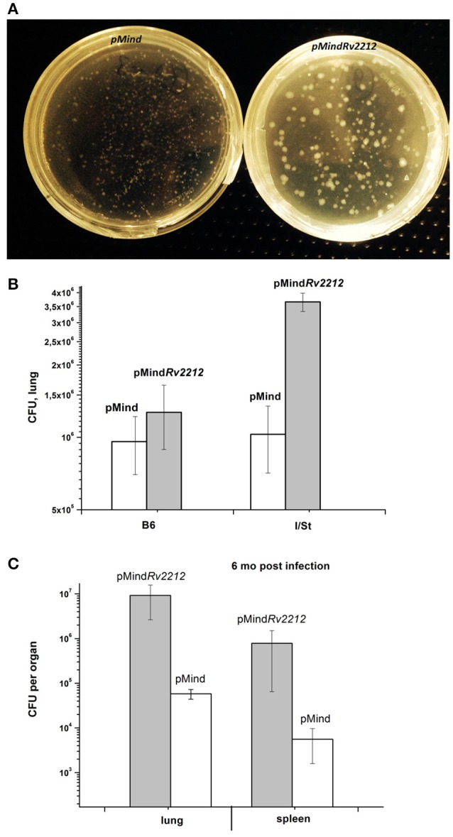
M. tuberculosis pMindRv2212 and pMind phenotypes expressed in vivo. (A) pMindRv2212 strain (left) isolated from lungs provided larger colonies compared to the control strain (right). Lungs of infected B6 mice were homogenized, plated on Dubos agar, kept at 37°C for 3 weeks and photographed. (B) Mice of I/St and B6 inbred strains were infected with 5 × 10⁶ CFUs of M. tuberculosis pMindRv2212 or pMind. At week three post-infection, lungs were homogenized and serial dilutions were plated on Dubo medium. The results of two experiments including 4 mice each (total N = 8) are expressed as mean ± SEM. Significant difference (P < 0.01, ANOVA) was observed between pMindRv2212-infected B6 and I/St mice. (C) At 6 months post-challenge, ~2 log differences (P < 0.001, ANOVA) were observed between multiplication of pMindRv2212 and pMind mycobacteria in lungs and spleens of B6 mice. The results are expressed as mean ± SEM (N = 6).
