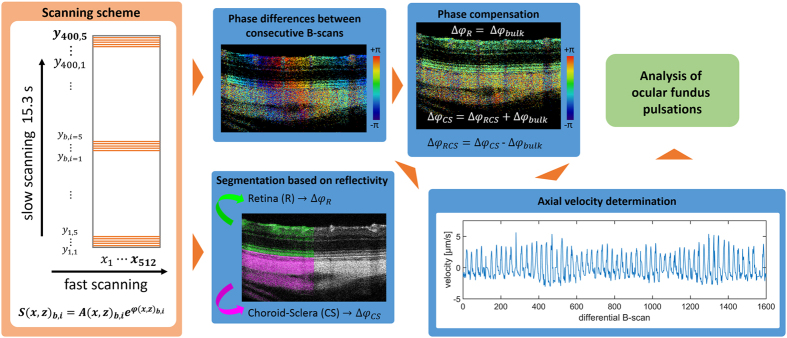
Figure 1.
Overview of the ocular fundus pulsation measurement. A repeated raster scanning pattern was used to perform phase-differencing between consecutive B-scans. After segmenting the retinal and chorioscleral region, bulk phase shifts (estimated in the retina) were subtracted from each B-scan and the remaining average phase differences between the retina and chorioscleral complex were calculated. The axial velocities of these deformations were subsequently used for a quantitative spectral analysis.

