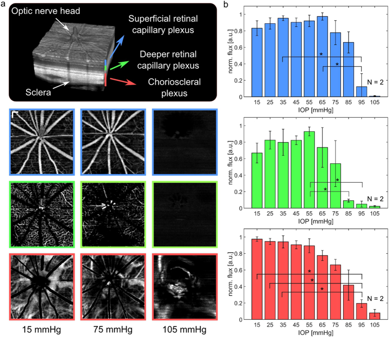
Figure 4.
Changes of the retinal perfusion in response to increased IOP. (a) En-face OCTA maximum-intensity-projections of three plexuses at three different IOP levels. Non-perfused patches in the deeper retinal capillary plexus were observed at increased IOP levels of 75 mmHg. While the retinal perfusion stopped at an IOP level of 105 mmHg, some major choroidal vessels were still present in the OCTA images. (scale bar = 150 μm) (b) Quantitative analysis of the flux in all three plexuses in four animals. The flux in the superficial retinal plexus dropped significantly in the range of 65 to 95 mmHg while the flux in the deeper retinal capillary plexus earlier decreased significantly between 55 mmHg and 85 mmHg. (*significant difference: p < 0.05).

