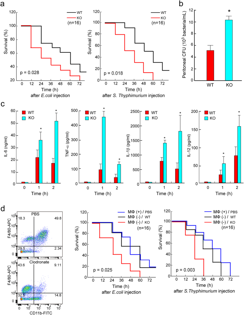
Figure 6.
TAGLN2 participates in host survival after bacterial peritonitis. (a) TAGLN2 −/− mice demonstrate reduced survival after bacterial peritoneal sepsis. Age-, sex-, and weight-matched wild-type (black line) and TAGLN2 −/− (red line) mice were injected i.p. with 5 × 10⁸ CFU of live E. coli (DH5α) or 5 × 10⁶ CFU of live S. typhimurium. Kaplan-Meier survival curves revealing the proportion of mice that are alive in each group as a function of time from the first injection of bacteria are shown. (b) The E. coli cell count in peritoneal lavage fluid from wild-type or TAGLN2 −/− mice 4 h after i.p. administration of 108 CFU/mouse of E. coli is shown as bar graph (n = 6). *P < 0.05 vs LPS-treated wild-type macrophages. (c) Cytokine (TNF-α, IL-1β, IL-6, and IL-12) levels in plasma at 0, 1, and 2 h after i.p. injection of 5 × 10⁶ CFU of live S. typhimurium into wild-type and TAGLN2 −/− mice. Data are the mean ± SEM; n = 8 per group. *i < 0.05 vs. wild-type mice at the same time point. (d) Flow cytometry plots of CD11b+, F4/80+ macrophages isolated from spleens at day 2 after clodronate liposomes treatment for depletion (n = 3 per group). Survival after transfer of wild-type and TAGLN2 −/− peritoneal macrophages in recipient wild-type mice with E. coli- or S. typhimurium-induced sepsis (n = 15/group).

