Figure 4.

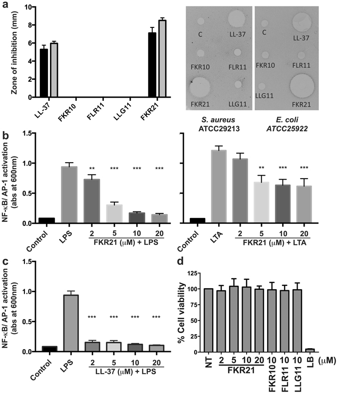
Antimicrobial and immune-modulatory effects of LL-37 and peptide fragments. (a) Determination of antimicrobial activity (using radial diffusion assay, RDA) of the peptide fragments (FKR10, FLR11, LLG11 and FKR21) and LL-37 (100 μM) against S. aureus ATCC29213 (black bars) and E. coli ATCC25922 (grey bars) (n = 6). The right panel illustrates two scanned examples of representative RDA gels visualizing the zones of clearance corresponding to the inhibitory effects of the peptides against S. aureus and E. coli after incubation at 37 °C for 18–24 h (C, control, buffer 10 mM Tris pH 7.4). (b) Evaluation of NF-κB/AP-1 activation in supernatants of THP1-X-Blue CD14 cells after stimulation with 100 ng/ml of E. coli LPS (left) or 1 μg/ml of S. aureus LTA (right) and increasing concentrations of FKR21. (c) Evaluation of NF-κB/AP-1 activation in supernatants of THP1-X-Blue CD14 cells after stimulation with 100 ng/ml of E. coli LPS and increasing concentrations of LL-37 are shown for comparison. (d) Cell viability of HaCaT keratinocytes was analysed using a MTT assay. The results are indicated as mean absorbance values after treatment by the LL-37 derived peptide fragments, which correspond to the amount of living cells, and are compared to the control representing non-treated (NT) cells. Lysis Buffer (LB) yielded 100% lysis of the cells, mean values and SD of at least three independent experiments are presented (**P < 0.01, ***P < 0.001).
