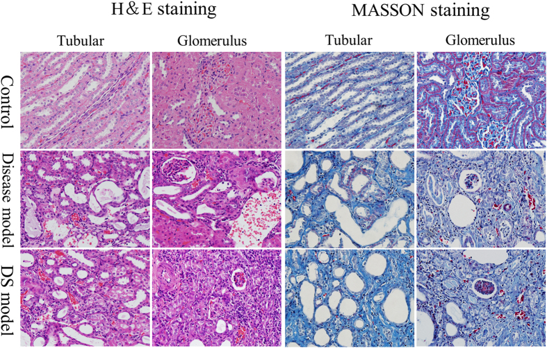
Figure 2.
H& E and Masson staining on kidney tissue of rats in control, disease and DS groups. H& E staining: rats in disease and DS groups were observed with renal cortex and medulla atrophied, glomerular sclerosis and atrophy, renal tubular expansion, structure diffusion, visible hyalinization, protein tube and a large number of cavity; Glomerular and capillary congestion and clear inflammatory cell infiltration was also observed. Masson staining: glomerular basement membrane and tubule interstitial fibers proliferated significantly in disease and DS groups of rats.

