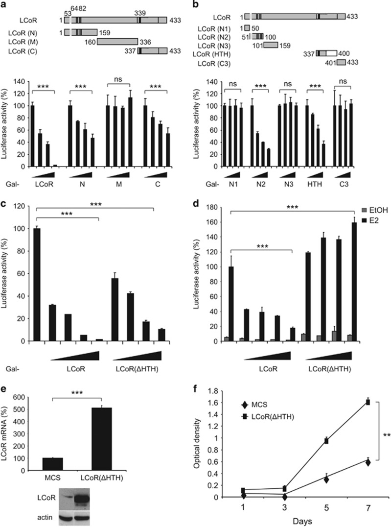
Figure 3.
The HTH domain has a major role in LCoR activity. (a, b) Upper panels. Schematic representation of LCoR fragments. Lower panels. COS7 cells were transiently transfected with pRL-CMV-renilla and L8G5 reporter plasmids, LexAVP16 and increasing doses of plasmids expressing the various LCoR variants fused to Gal4DBD. Luciferase values were expressed as described in Figure 2a. (c) T47D cells were transiently transfected with pRL-CMV-renilla and L8G5 reporter plasmids, LexAVP16 and increasing doses of Gal4DBD-LCoR or -LCoR(ΔHTH) expression plasmids. Luciferase values were normalized as described in Figure 2a. (d) T47D cells were transiently transfected with pEBL+ (encoding ERE-driven luciferase), pRL-CMV-renilla as internal control and increasing doses of p3XFlag-LCoR or p3XFlag-LCoR(ΔHTH) expression vectors. Cell treatment and data normalization were as in Figure 2b. (e) T47D cells were stably transfected with LCoR(ΔHTH). LCoR(ΔHTH) mRNA and protein expression were checked by real-time PCR (upper panel) and western blotting with an anti-Flag and an anti-actin antibody (lower panels), respectively. (f) Cell proliferation was measured as described in Figure 2d. Statistical analyses were performed using the Mann–Whitney test. NS, not significant; **P<0.01; ***P<0.001.

