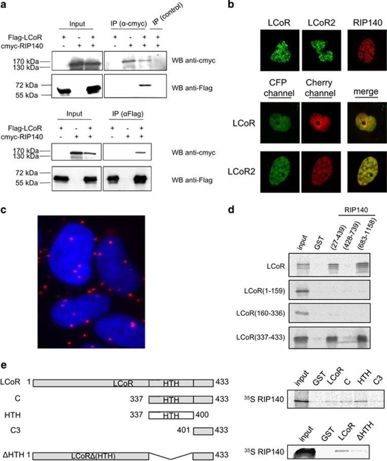
Figure 6.
LCoR interacts with RIP140. (a) LCoR and RIP140 coimmunoprecipitate. COS7 cells were transfected with Flag-LCoR and/or c-Myc-RIP140 expression plasmids, as indicated. Two days after transfection, whole-cell protein lysates were immunoprecipitated with the anti-c-Myc, the anti-Gal4DBD antibody (control) (upper panels) or the anti-Flag antibody (lower panels) and analyzed by western blotting using anti-c-Myc or anti-Flag antibodies. An aliquot of each extract was also immunoblotted to evaluate the expression levels of LCoR and RIP140 (input). (b) COS7 cells were transfected with CFP-LCoR, CFP-LCoR2 and/or Cherry-RIP140 expression plasmids, as indicated. Two days after transfection, cells are analyzed as in Figure 1c. (c) In situ proximity ligation assay between LCoR and RIP140 in MCF7 cells using anti-RIP140 and anti-LCoR antibodies diluted in phosphate-buffered saline–1% bovine serum albumin. (d) GST pull-down assays were carried out using bacterially expressed GST and GST-RIP140 fragments and 35S-labeled LCoR and LCoR deletion mutants. (e) Schematic representation of full-length and LCoR deletion mutants (left). GST pull-down assays were carried out as in d with GST and the indicated GST-LCoR fragments and 35S-labeled RIP140 (right).

