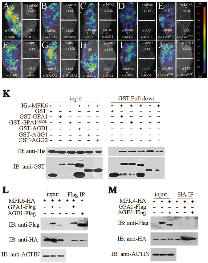
Figure 1.

AGB1, GPA1, AGG1 and AGG2 interact with MPK6 and MPK3. (A–J) Tobacco leaves co-infiltrated with agrobacterium containing 35S-driven split luciferase (LUC) constructs as indicated were photographed with a charge-coupled device camera. Each image is representative of three images in three independent experiments. (A–E) MPK6-nLUC interacts with cLUC-AGB1 (A), cLUC-WtGPA1 (B), cLUC-AGG1(C), cLUC-AGG2 (D), cLUC-GPA1Q222L (E), respectively. (F–J) MPK3-nLUC interacts with cLUC-AGB1 (F), cLUC-WtGPA1 (G), cLUC-AGG1 (H), cLUC-AGG2 (I), cLUC-GPA1Q222L (J), respectively. The pseudocolor bar shows the relative range of luminescence intensity in images. Pull-down assay shows that AGB1, WtGPA1, GPA1Q222L, AGG1 and AGG2 interact with MPK6 (K), respectively. AGB1 and GPA1 interact with MPK6 (L), but not MPK4 (M) by Co-IP assay. Full blots are shown in Supplemental Data.
