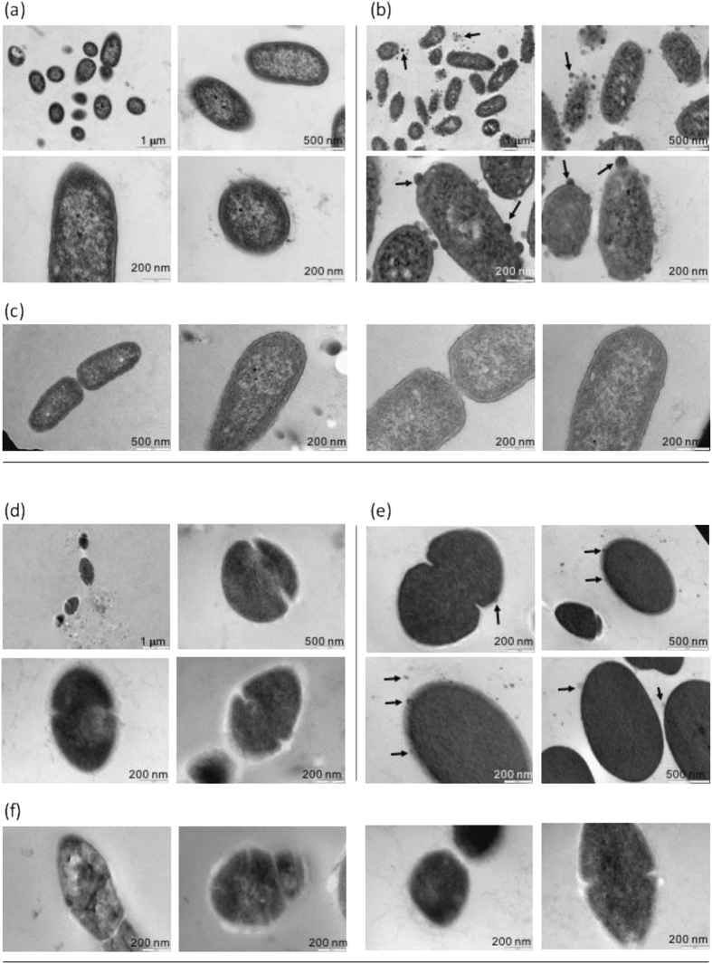
Figure 2.
Transmission Electron Microscope images of P. aeruginosa ATCC 10145 and S. thermophilus DSM 20617 T before and after exposure to chlorhexidine and promysalin. (a) P. aeruginosa cell not exposed and (b) exposed to chlorhexidine (100 µg/ml) or (c) to promysalin (100 µg/ml). (d) S. thermophilus cell not exposed and (e) exposed to chlorhexidine (100 µg/ml) or (f) to promysalin (100 µg/ml). Black arrows indicate the membrane protrusions in cell exposed to chlorhexidine.

