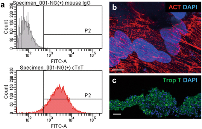
Figure 1.

Characterization of hiPS-CMs and hiPS-CM cell sheet. (a) Expression of cardiac troponin T (cTNT) after differentiation and purification of hiPS-CMs was determined by flow cytometry anaysis. (b) After differentiation and purification, sarcomere structure was visualized by sarcomeric alpha actinin staining in hiPS-CM. (c) Immunostaining of the hiPS-CM cell sheet with cTNT antibody (green). The cell nuclei were counterstained with 4′,6-diamidino-2-phenylindole (DAPI; blue). Scale bar, 10 μm in (b) and 100 μm in (c).
