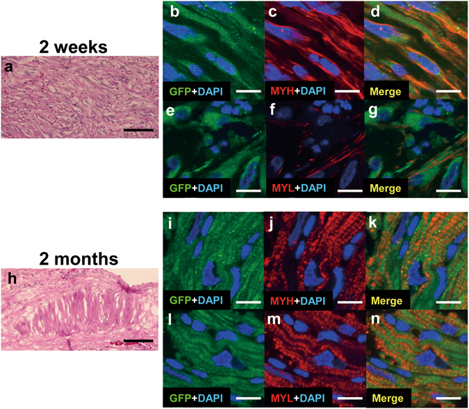
Figure 8.

The EGFP labeled hiPS-CMs after transplantation with the omentum. (a–g) Detection of EGFP-labeled hiPS-CMs 2 weeks after transplantation; representative photomicrographs showing HE stain (a), GFP in green (b,e), myosin heavy chain (MYH) and myosin light chain-2 (MYL) in red (c,f). (h–n) Detection of EGFP-labeled hiPS-CMs 2 months after transplantation; representative photomicrographs showing HE stain (h), GFP in green (i,l), and MYH and MYL in red (j,m). The nuclei were stained with DAPI in blue (b–g,i–n). Merged images are shown in (d,g,k,n). Bar = 100 μm in (a,h), 10 μm in (b–g,i–n).
