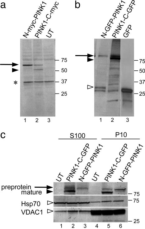
Fig. 2.
Processing of PINK1 protein in mammalian cells. COS-7 cells were transfected with myc-tagged (a) or GFP-tagged (b) constructs. N-terminal (lane 1) and C-terminal (lane 2) tagged versions were used, and either untransfected cells (lane 3 in a) or GFP-transfected cells (lane 3 in b) were included as controls. Arrows show preprotein (before cleavage of the N terminus), and filled arrowhead indicates mature peptide. Open arrowhead in the GFP constructs shows a possible N-terminal fragment (see Results); *, cross-reactivity seen with myc monoclonal in untransfected cells. (c) Subcellular fractionation. COS-7 cells were transfected with C-terminal (lanes 2 and 5) or N-terminal (lanes 3 and 6) GFP-tagged PINK1 or untransfected (lanes 1 and 4) and cell lysates separated into a 10,000 × g pellet (P10, lanes 4–6) and 100,000 × g supernatant (S100, lanes 1–3) fractions. Blot was reprobed with antibodies to Hsp70 and VDAC1 in Middle and Bottom, respectively (open arrowheads). Data are representative of more than two experiments for each construct. Markers on the right of all blots are in kilodaltons.

