
Figure 3. Distal axon-derived TrkA endosomes in dendrites are signaling competent and localized in close proximity to PSDs.
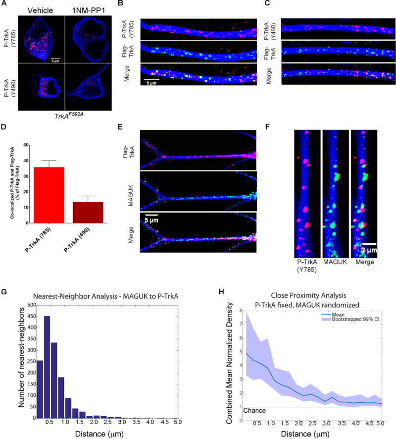
A. Images of TrkAF592A cell bodies immunostained for P-TrkA (Y785) puncta in both vehicle (top left) and 1NMPP1 (top right) conditions, as well as cell bodies immunostained for P-TrkA (Y490) puncta in both vehicle (bottom left) and 1NMPP1 (bottom right) conditions, showing specificity of P-TrkA antibodies. B–H represent data 3–6 hours after NGF application to the distal axon compartment. B, C. Images illustrating co-localization of retrograde Flag-TrkA endosomes and P-TrkA (Y785) (B) and Y490 (C) in dendrites. C is same scale as B. D. Quantification of co-localization between retrogradely trafficked Flag-TrkA endosomes and P-TrkA (Y785) and P-TrkA (Y490) (n=3). E. Images of distal axon-derived Flag-TrkA endosomes (top) in close proximity to the PSD protein MAGUK (middle) in dendrites of sympathetic neurons. F. High magnification image of P-TrkA puncta (left) within dendrites in close proximity to MAGUK puncta (middle). G. Histogram of nearest neighbor analysis of nearest MAGUK puncta to P-TrkA (Y785) puncta. H. Population level analysis comparing the proximity of P-TrkA (Y785) and MAGUK puncta to that expected by chance, represented on the y-axis as 1. Transparent band shows 99% confidence intervals determined by bootstrapping. G,H n=22 dendrites, 4 chambers, 2 independent experiments. A–F MAP2 ICC is in blue. Data are presented as mean ± SEM. See also Figure S2.