
Figure 4. Distal axon-derived, signaling competent TrkA endosomes are localized within sympathetic neuron dendrites in vivo.
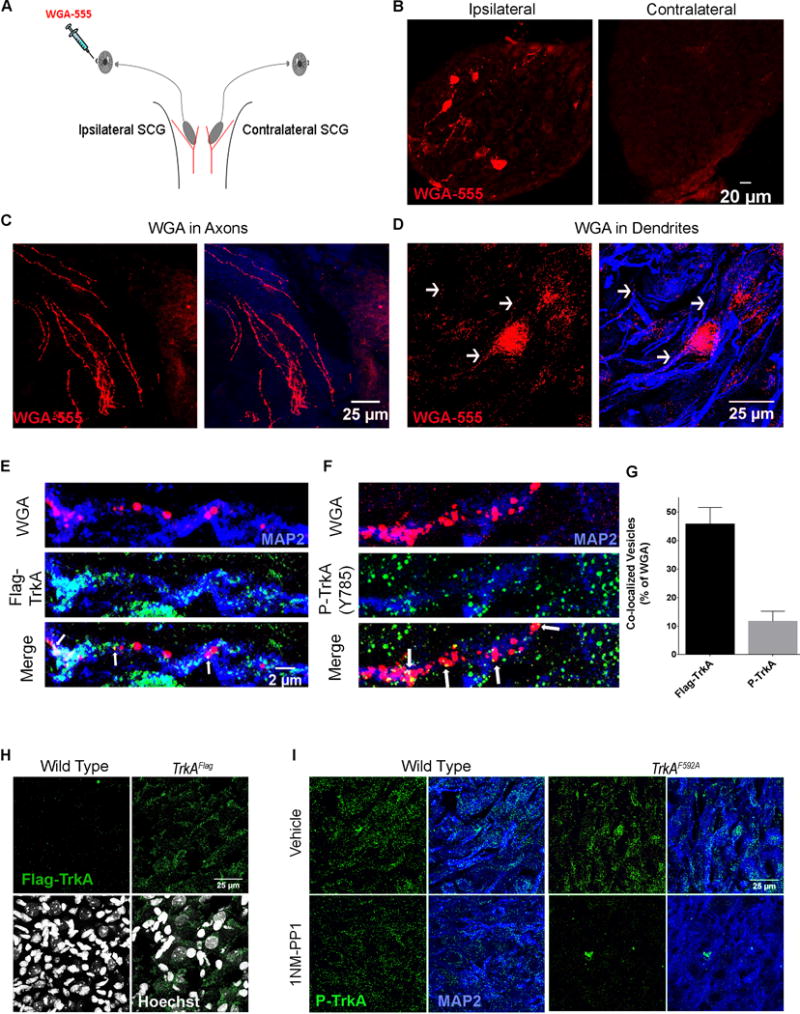
A. Schematic of the assay used to track retrograde vesicle transport from the ipsilateral sympathetic target field to postganglionic neuron CBs and dendrites. WGA-555 injected into the anterior chamber of the eye is endocytosed by sympathetic neuron distal axons and transported retrogradely to the CBs and dendrites of sympathetic neurons residing in the SCG; located where the carotid artery branches into internal and external branches (red). B. Specific WGA-555 labeling of neurons in the ipsilateral ganglion (left) but not the contralateral (right). C. WGA-555 vesicles in sympathetic axons. D. WGA-555 vesicles in MAP2+ dendrites. E. Retrogradely trafficked WGA vesicles are co-localized with Flag-TrkA puncta in dendrites 16 hours post injection. F. Retrogradely trafficked WGA vesicles are co-localized with P-TrkA puncta in dendrites 16 hours post injection. E is same scale as F. G. Quantification of E and F, n=3 animals. H. 5 μm sections of SCG tissue immunostained for Flag-TrkA and labeled with Hoescht (bottom), showing specific Flag puncta not in wild-type (left) but only in TrkAFlag (right) animals. I. 3 μm sections of SCG tissue immunostained for P-TrkA (Y785) and MAP2 of both WT (left) and TrkAF592A (right) animals treated with IP injections of either vehicle (top) or 1NMPP1 (bottom). There is specific reduction of P-TrkA puncta in 1NMPP1 treated TrkAF592A but not control animals. Data are presented as mean ± SEM. See also Figure S3.