
Figure 5. TrkA signaling endosomes are found in dendrites and in close proximity to synapses throughout development in vivo.
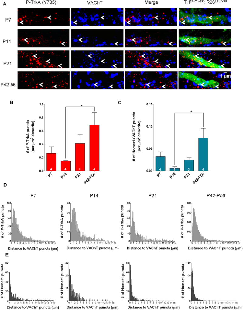
A. Dendrites of TH2A-CreER;R26LSL-YFP (Ai3) sparsely labeled cells in the SCG at developmental time-points P7, P14, P21 and P42–P56 immunostained for P-TrkA (Y785) (left) and VAChT (left middle). Arrowheads denote P-TrkA (Y785) puncta found in dendrites close to VAChT puncta. B. Quantification of the number of P-TrkA puncta per μm2 of labeled dendrite. One way ANOVA F (3, 11) = 3.845 P<0.05 post hoc Tukey’s multiple comparisons’ test: *p<0.05. C. Quantification of the number of co-localized Homer1 and VAChT puncta per μm2 of labeled dendrite. One way ANOVA F (3, 11) = 5.147 P<0.05 post hoc Tukey’s multiple comparisons’ test: *p<0.05. D. Histograms of the distance between P-TrkA puncta and the nearest VAChT puncta within labeled dendrites. E. Histograms of the distance between Homer1 puncta and their nearest VAChT puncta within labeled dendrites. D, E bin size is 0.1 μm. Data are presented as mean ± SEM. See also Figure S4.