
Figure 7. Distal axon-derived TrkA signals locally, within dendrites, to maintain PSDs in sympathetic neurons.
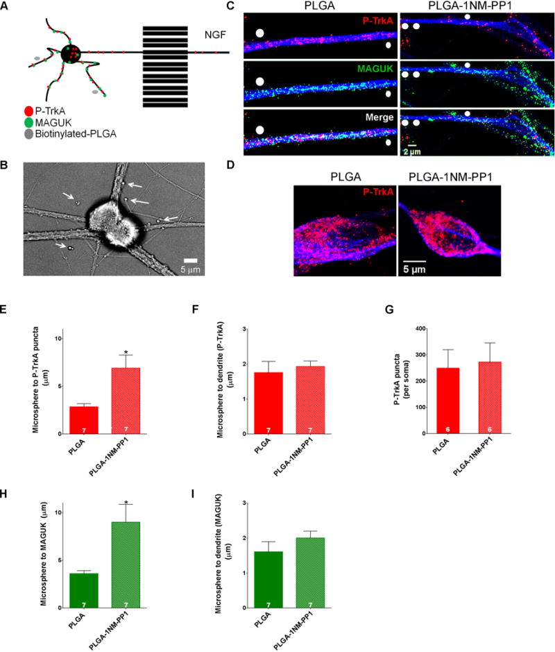
A. Biotinylated-PLGA-1NMPP1 microspheres were applied to the somatodendritic compartment of DIV 14 compartmentalized TrkAF592A sympathetic neurons cultured on coverslips pre-coated with streptavidin while NGF was applied to distal axons to achieve localized TrkA kinase inhibition pockets adjacent to dendrites. B. Bright field image of SCG neurons. Arrows denote PLGA-1NMPP1 microspheres. C. P-TrkA and MAGUK puncta in dendrites of neurons treated with PLGA (left) or PLGA-1NMPP1 (right) microspheres. White circles are drawn over microspheres. D. P-TrkA puncta in soma of neurons treated with PLGA (left) or PLGA-1NMPP1 (right) microspheres, quantified in G. E and H. Quantification of the distance between PLGA or PLGA-1NMPP1 microspheres and the nearest P-TrkA (D) or MAGUK (G) puncta; Welch’s t-test*p<0.05. F and I. Quantification of the distance between each PLGA microsphere and dendrite mask per experiment. Data are presented as mean ± SEM. See also Figure S6.