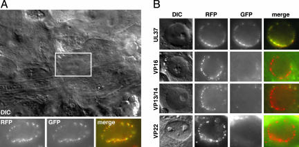
Fig. 4.
Accumulation of capsids and tegument proteins at the nuclear rims of infected sensory neurons after retrograde transport. (A) Differential interference-contrast image of the center of a DRG explant infected with the mRFP1-VP26–GFP-VP1/2 virus (2 hpi). The fluorescence signals associated with the nucleus of a sensory neuron (boxed) are shown below. (B) Nuclear rim images of the remaining dual-fluorescent viruses, carried out as in A. Labels at the left indicate the GFP fusion present in each infection.

