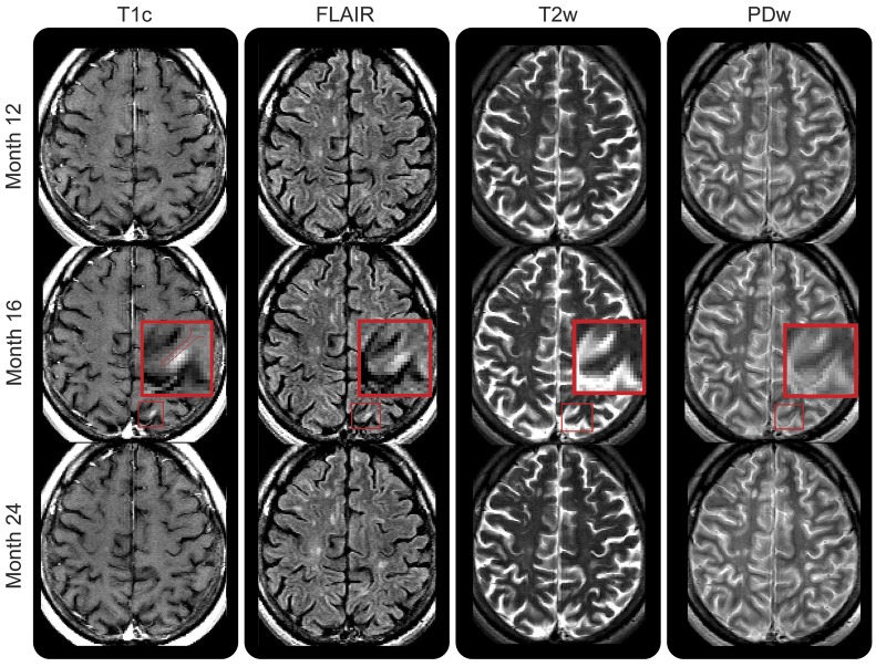
Figure 3. Gadolinium-enhancing intracortical lesion with resolving T2-weighted hyperintensity.
Small enhancing lesion affecting the cortex (red box indicates the magnified area) that appears at month 16 on contrast-enhanced T1-weighted imaging, fluid-attenuated inversion recovery (FLAIR), and T2/proton density (PD)–weighted imaging, and resolves by month 24. Enhancement appears limited to the cortex, but due to the resolution of the postgadolinium T1-weighted image, the possibility that the enhancement involves the white matter cannot be excluded. PDw = proton density-weighted imaging; T1c = T1-weighted image postgadolinium injection; T2w = T2-weighted imaging.

