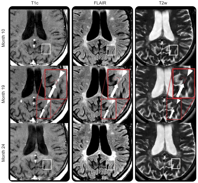
Figure 4. Cortical and subcortical gadolinium-enhancing lesions.
Enhancing lesion affecting the cortex (white arrow) next to an area of enhancement in the white matter (WM) (white arrowhead). The cortical enhancement appears distinct from the WM enhancement. Both areas of enhancement are observed first at month 19 in an area of normal-appearing tissue (white box on month 10 scan: first row), and regress, leaving a subtle hyperintense signal on T2-weighted imaging (white box on month 24 scan: last row). FLAIR = fluid-attenuated inversion recovery; T1c = T1-weighted image postgadolinium injection; T2w = T2-weighted imaging.

