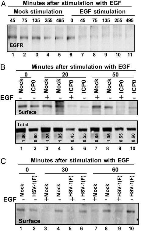
Fig. 5.
Cell surface and total EGFR levels are affected by ICP0 or by wild-type virus. (A) Effect of EGF on EGFR on cell surface. Replicate HEK293 cells grown in T-25 flasks were stimulated or mock-stimulated with 100 ng/ml EGF at time intervals shown. EGFR on cell surfaces was biotinylated and detected as described in Materials and Methods. (B) EGFR in cells transfected with ICP0. Replicate cultures of HEK293 cell flasks were transfected with EGFR and MTS1-ICP0 or the same amount of empty vector MTS1. At 12 h after transfection, the cultures were replenished with serum-free medium. At 36 h after transfection, the cells were mock-stimulated or exposed to EGF for 20 or 50 min. The cell surface levels of EGFR were assayed as described above (Upper). An aliquot of the total cell lysate removed before immunoprecipitation served to measure the level of EGFR in the total cell lysate (Lower). (C) EGFR in cells infected with HSV-1(F). Replicate cultures of HEp-2 cells were incubated in serum-free medium for 24 h, then mock-infected or infected with HSV-1(F). At 4 h after infection, the cells were mock-stimulated or exposed to EGF for 30 or 60 min. The cell surface levels of EGFR were assayed as detected above.

