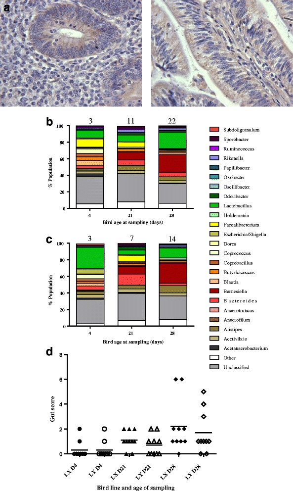
Fig. 6.

AvBD1 IHC and caecal microbiota analyses of LX and LY birds. IHC analyses to show epithelial localisation of AvBD1 in caecal tissues of Day 4 LX birds using AvBD1 polyclonal antibody diluted 1:250 and peroxidase staining (×400 magnification) (a). Caecal microbiota profiles of LX (b) and LY (c) birds (two independent trials using two different hatches) presented as relative abundance of bacterial genus (% microbial population). Corresponding total gut health scores/sampled group at each time point is shown above each column. (d) individual gut health data focussing on redness, watery digesta and gut tone where 0 (normal), 1 (some abnormalities) or 2 (very abnormal), and poorest guts scoring 6
