
Fig. 5.
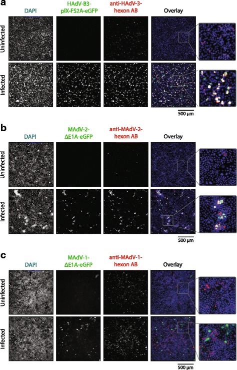
Use of hexon fragment antibodies in immunofluorescence analysis of GFP-reporter virus infected cells. a Human A549 cells were infected with replication-competent HAdV-3-pIX-FS2A-eGFP at an MOI of 1 and cells were fixed and stained one day post infection with the HAdV-B3 hexon Ab. Co-localization of a HAdV-B3 hexon specific signal with the GFP signal was observed in the overlays. b Mouse 3T6 cells were infected with 100 geq vp of MAdV-2-ΔE1A-eGFP and cells were fixed and stained five days post infection with the MAdV-2 hexon Ab. Co-localization of the MAdV-2 hexon signal with GFP was observed in channel overlays. c Mouse 3T6 cells were infected with 100 geq vp of MAdV-1-ΔE1A-eGFP and cells were fixed and stained five days post infection with the MAdV-1 hexon Ab. No co-localization of GFP and hexon signal was observed. DAPI staining is highlighted in blue, the GFP signal is highlighted in green and the hexon antibody signal is highlighted in red