
Fig. 6.
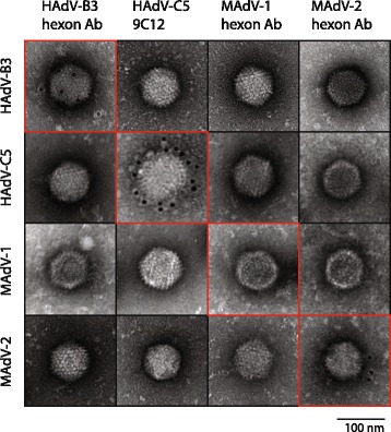
Transmission electron microscope analysis of immunogold-labeled viral capsids. Purified HAdV-B3, HAdV-C5, MAdV-1 and MAdV-2 were first incubated with the indicated rabbit hexon antibodies for HAdV-B3, MAdV-1, MAdV-2, or the monoclonal mouse 9C12 antibody specific for HAdV-C5, respectively. Following staining with colloidal 10 nm gold-labeled anti-rabbit or anti-mouse antibodies, uranyl-acetate staining was performed. TEM images were acquired at 135 000× magnification